Welcome! This blog contains research & information on lifestyle, nutrition and health for those with MS, as well as continuing information on the understanding of the endothelium and heart-brain connection. This blog is informative only--all medical decisions should be discussed with your own physicians.

The posts are searchable---simply type in your topic of interest in the search box at the top left.

Almost all of MS research is initiated and funded by pharmaceutical companies. This maintains the EAE mouse model and the auto-immune paradigm of MS, and continues the 20 billion dollar a year MS treatment industry. But as we learn more about slowed blood flow, gray matter atrophy, and environmental links to MS progression and disability--all things the current drugs do not address--we're discovering more about how to help those with MS.

To learn how this journey began, read my first post from August, 2009. Be well! Joan

Saturday, June 27, 2015

Pictures. Worth some words.

I write a lot.  Today I just wanted to share some images, because these pictures speak loudly and can amplify recent blog posts.  Some neurologists are saying there's no way the newly discovered lymphatic vessels and CCSVI are related.  But they are wrong.  Just as they have been wrong about the brain's "immune privilege" and the EAE mouse model of MS.
http://ccsviinms.blogspot.com/2015/06/rewrite-textbooks.html

The following images are all of the venous and lymphatic drainage of the brain, from the back of the head.

This is an MRI picture from the Kipnis Lab at the University of Virginia, of the newly discovered lymphatic vessels, shown with red dye.  These were also found in human cadaver tissue.  These newly discovered vessels directly follow the path of the brain's draining veins, shown with green dye.


This is an illustration of the veins which drain the human and mammalian brains.  Cortical veins at the top of the head lead into the superior sagittal sinus.  They branch off on either side, forming the transverse sinus, where they eventually head south into the jugular veins. See how nice and open they appear? Lymph vessels follow this system.  Lymph vessels drain lymph carrying immune cells, metabolites, proteins and toxins from the brain into jugular veins.  You can see how the mouse brain image above matches this.


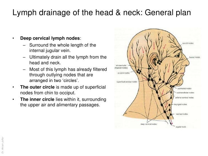
The image below shows the location of the cervical lymph nodes.  All of the lymph collected in the newly discovered vessels flows alongside the veins and back to the cervical lymph nodes. Notice that they are situated alongside the jugular vein.


AND finally, this is an MRV image of my husband Jeff's brain draining veins. Notice the straight cortical veins, leading down into the transverse sinus, where they branch off. And then....what the what? His sigmoid sinus and jugular foramen are pinched off, creating a mess of curly collaterals beneath.  His brain's blood flow and lymph were all slowed to a stall, waiting to be being drained.  He was sick, tired, had over 20 cerebral lesions, and had just received an MS diagnosis.


His sigmoid sinus and jugular veins have since been stented, and he now has normalized flow going through his jugular veins. The collaterals disappeared. How has this impacted his brain health? Well, he hasn't had any more MS lesions and his gray matter atrophy reversed.   He's running around, always on the go, very thankfully still driving me crazy. Yes, this is all anecdotal.  But sigmoid stenting is an approved treatment for intracranial hypertension.  It's not vodoo.  It improves cerebrospinal fluid and blood flow, and helps people heal.  And now we know, this is where lymph flows, as well.

I would contend that this is something SOMEBODY might like to study. All it would take would be to block the mouse veins at the sigmoid sinus, insert the tracer dye into the lymphatic vessels, and watch what happens to the drainage of lymph.   I'll bet you it isn't good.

The time is now,

Joan


Sunday, June 21, 2015

Rewrite the textbooks

UPDATE: October 2017  
Confirmation of the Kipnis Lab and University of Helsinki finding in humans.  
Researchers at NIH see the actual lymphatic vessels in the human brain. 

“We literally watched people’s brains drain fluid into these vessels,” said Daniel S. Reich, M.D., Ph.D., senior investigator at the NIH’s National Institute of Neurological Disorders and Stroke (NINDS) and the senior author of the study published online in eLife(link is external). “We hope that our results provide new insights to a variety of neurological disorders.”   link

                                                   *******************

The Journal of Experimental Medicine said it best in their recent tweet:

"The CNS is considered an organ devoid of lymphatic vasculature." Ed. note: "NOT"

Not. Decades of medical theory which considered the brain as different from other organs of the body and without lymphatic drainage system were, well, simply wrong.

The University of Helsinki has independently published a paper in the Journal of Experimental Medicine which confirms the University of Virginia's paper on lymph vessels published just two weeks ago. Both universities have found:
A dural lymphatic vascular system that drains brain interstitial fluid and macromolecules

How have researchers had it SO WRONG for SO LONG?
If you're like me, you figured that neuroscientists had looked long and hard for any signs of lymphatic drainage surrounding the brain, and had proven that it simply didn't exist, and that the brain was indeed immune privileged, and separate from the rest of the body's peripheral immune system.

“Any neuroscience textbook that has ever been written will say that the central nervous system is devoid of a lymphatic system and that is one of the reasons the brain is immune privileged,” Louveau said. “When we started our project, our question was if there are so many immune cells surrounding the brain, how do they traffic there? By addressing this question we found vessels that weren’t supposed to exist. They were very well hidden and we think that is why it took so long to discover them.”
link

In fact, the whole concept of immune privilege of the brain and lack of lymphatic drainage was created 70 years ago and hasn't changed much in ensuing years.   This was around the same time EAE was created as the animal model for MS.  Why hasn't there been more exploration until recently?   I believe one reason is because the old theories have made people very rich.

Let's learn the history:
The theory of  immune privilege was invented to explain why foreign tissue grafts placed on brain tissue didn't cause an immediate immune reaction, as similar grafts did in other parts of the body, like the skin.  It was believed that antigens in the brain were concealed from the immune system by the blood brain barrier.  That's why it was assumed that when immune cells showed up in the brain--if they weren't there fighting an infection or as an inflammatory reaction after a stroke---there could only be one explanation--it was some sort of "auto-immune"and destructive inflammatory reaction.  

This theory of immune privilege was developed in the 1940s and became the foundation of transplantation immunology---or the reason why we need to block the immune system when patients receive a donated organ or a skin graft.  The body's immune response needs to be turned off, so that it will not reject the foreign tissue.  This process was discovered by Sir Peter Medawar, who wanted to understand how to help skin grafts survive.


Sir Peter Medawar, whose experiments in the 1940s established the basic rules of transplantation immunology [2], placed skin and or other types of grafts in the anterior chamber of the eye and the brain [3]. Observing that many of these grafts, unexpectedly, survived for prolonged intervals of time, he invented the term ‘immune privilege‘ to refer to the unexpected and prolonged acceptance of solid tissue grafts at specialized sites in the body. At the time, it was believed that the brain and eye lacked lymphatic drainage pathways, and that both organs resided behind stringent blood-tissue barriers. In light of this knowledge, Medawar proposed that immune privilege results from ‘immunologic ignorance’ because, on the one hand, in the absence of lymph pathways antigens could not escape from the eye, and on the other hand, immune effectors in the blood could not pass the vascular barrier to enter the sites where antigen resides. He speculated that antigenic material placed in privileged sites is sequestered from the immune system, and that the system remains unaware of the existence of grafts in privileged sites. link

This speculation on immune privilege has continued on for seven decades, mostly due to the fact that no one could find any lymphatic drainage pathways or understand how or why immune cells might get through the blood brain barrier.  

Not coincidently, the EAE model for MS was developed around the same time as the theory of immune privilege, as an example of how t-cells could break through the blood brain barrier and attack the brain in an auto-immune reaction.  It has been a prevailing theory in MS treatment development, which has also never been proven.  Another 70 year old theory which has lead to disastrous assumptions about the brain.


The presence of mild scant lymphocytic infiltrates in the demyelinating lesions has been generally interpreted as the evidence of an inflammatory autoimmune process. Because specific T-cell mediated autoimmunity can be reproduced in animals after myelin protein sensitisation (Experimental Allergic Encephalo- myelitis (EAE)) it has been assumed (but never proven) that a similar T-cell driven immune mechanism is responsible for demyelination in MS. 

The acceptance of EAE as a model for MS is an unfortunate error that has its basis on faith rather than science. Whilst EAE is a good example of an experimental organ-specific autoimmune disorder in animals, it cannot be accepted as a model for MS for a wide variety of reasons. This is particularly important in relation to the development of MS pharmacotherapy. We have analysed the literature on immune-modifying therapy in MS and it is clear that none of these agents can qualify as a candidate therapy under scrutiny.
link


The brain's relationship to the peripheral immune system is obviously much more communicative and complex than previously imagined.  The discovery of lymphatic vessels by neuroimmunologists means we need a do-over in MS research, as this science writer comments:

This data suggests that brain immune surveillance communicates with the immune system and can generate adaptive immune responses. The authors infer that previously characterized glymphatic washing of the brain likely connects to the lymphatic system; a testable hypothesis. The authors further suggest that this system will change the way we think about neurological disorders such as multiple sclerosis and Alzheimer’s.

The fact that MS has no disease-specific immune target,  no specific antigen, and does not look like EAE in mice, should give us all pause.   There has never been any concrete, scientific proof that MS is an autoimmune disease.  Just a theory,  which has been used to create a 20 billon dollar a year industry.  And that theory has completely unravelled.  The question is, will MS researchers let go of pharma money, in order to pursue the true etiology of MS?

If the brain requires immune cells for neuroprotection, stem cell regeneration and plasticity, than what are we doing suppressing the immune system with MS drug therapies?  What if this immune reaction is, as Dr. Michal Schwartz's research has shown, "protective", and should be modified, not stopped?  (notice that this paper was co-authored by Dr. Kipnis, a PhD graduate of the Weizmann Institute of Science, who now heads up the Kipnis Lab at the Univeristy of Virginia where these lymphatic vessels were discovered.)

For decades, several axioms have prevailed with respect to the relationships between the CNS and circulating immune cells. Specifically, immune cell entry was largely considered to be pathological or to mark the beginning of pathology within the brain. Moreover, local inflammation associated with neurodegenerative diseases such Alzheimer's disease or amyotrophic lateral sclerosis, were considered similar in their etiology to inflammatory diseases, such as remitting relapsing-multiple sclerosis. The ensuing confusion reflected a lack of awareness that the etiology of the disease as well as the origin of the immune cells determines the nature of the inflammatory response, and that inflammation resolution is an active cellular process. The last two decades have seen a revolution in these prevailing dogmas, with a significant contribution made by the authors. Microglia and infiltrating monocyte-derived macrophages are now known to be functionally distinct and of separate origin. Innate and adaptive immune cells are now known to have protective/healing properties in the CNS, as long as their activity is regulated, and their recruitment is well controlled; their role is appreciated in maintenance of brain plasticity in health, aging, and chronic neurodevelopmental and neurodegenerative diseases.
link


Israel Steiner, a neurologist at the Hadassah University Hospital in Jerusalem, agrees that EAE has blocked "effective progress" for decades. He thinks alternative theories should be put to the test. "I definitely believe it's high time to reconsider the entire field. It has not led us into understanding the disease or to a better therapy for patients," he says. 
"Many people in the community who do not have a vested interest in the autoimmune hypothesis share my views, but I'm not sure they would like to step out."  
New Scientist vol 176 issue 2369 - 16 November 2002, page 12 

Dr. Steiner would later publish the paper:  Experimental allergic encephalomyelitis: A misleading model of multiple sclerosis  link


The MS neurologists with "vested interest in the autoimmune hypothesis" were the very same ones who shut down CCSVI research before it could even begin, or be done correctly.  They were the first to say that veins would have nothing to do with MS, that it was an autoimmune disease, that the brain's drainage system was unimportant.  They created the narrative that those of us pleading for collaboration with Dr. Paolo Zamboni and the International Society of Neurovascular Disease (www.isnvd.org) were wackos, a fringe element, a Facebook/YouTube phenomena that would pass.  

But they were wrong.  The brain's venous system is most certainly important for efficient drainage of CSF, blood and now, lymph.  And the brain's immune system appears to be intimately tied to venous flow of the brain via newly discovered lymph vessels.  link 

In fact, the two large lymph vessels which collect the lymph drainage from the brain, head and neck can enter the blood stream at the internal jugular vein, the subclavian vein, or the junction of these two veins, called the venous angle.  Lymph relies on the low pressure venous system for drainage. To say that this discovery has "nothing to do with CCSVI" is to blatantly ignore physiology.  For patients, or neurologists, who would like to learn more about lymphatics:  link

The writing is on the wall, in medical journals, blogs and even twitter.
And we are all watching, reading, and waiting.

Time to rewrite the textbooks and help people heal,

Joan


Tuesday, June 9, 2015

Dr. Michal Schwartz was right.

The last hundred years or so, all of neurological disease research has maintained that any immune cells which showed up in the brain were there for one of three reasons:  to protect the brain against infection, in an autoimmune reaction (as in MS) or during an inflammatory attack (as occurs after stroke or ischemia.)  But immune cells as part of a healthy, functioning brain?  Absolutely not!

MS researchers have continued this dogmatic narrative, based on the "successful" development of EAE and immune ablating and suppressing drugs.  They maintain that the brain is immune privileged, and the blood brain barrier is meant to keep immune cells out.  And they've been suppressing the heck out of MS patients' immune cells ever since.

Recent research from the University of Virginia and the Kipnis Lab has called all of this into question.  It now appears that a healthy immune system is connected to the brain,  Exactly as it is in all other organs, via lymphatic vessels which drain into veins.

But even before the U of V publication, there has been one researcher who has single-handedly challenged the dogma on the immune privileged brain.  She has asserted the need for immune cells in our central nervous system.   She has been ridiculed, mocked and ignored--because she has stated that immune cells are supposed to be in the brain to aid, repair and rebuild.  She has published on the premise that immune cells are neuroprotective.
link

Dr. Michal Schwartz of the Weizmann Institute of Science has questioned whether immune ablation and suppression  in multiple sclerosis drugs was the correct approach.  Her research showed that it was essential not to completely stop the immune cells entering the MS brain, but to retrain the cells:  immune modulation, rather than immune suppression.  She continued to publish that immune suppressing drugs had fallen short, that MS continued to progress.  She thought it was impossible that the brain would have given up its ability to be assisted by the immune system.  It simply did not make sense to her.  Why would our most important organ not need the immune cells?
link

Immune ablation and suppression never made sense to me.  Subsequently, I've been following her research ever since Jeff was diagnosed with MS in 2007.  And that's why Jeff has only ever been on Copaxone, which was developed at the Weizmann Institute.  Copaxone retrains the immune cells thought to be causing damage, and leaves the others intact.

Dr. Schwartz likes to quote Abraham Lincoln--
"If you are doing any revolution, do not try to convince your opponents,
if you are right, you don't need it.  If you are wrong, it will not help you."

Dr. Schwartz has been quietly conducting her revolution in Israel, publishing her research in medical journals, and speaking out on the absolute necessity of immune cells in the brain.  As she says, "A healthy mind depends on a healthy immune system."  These so-called lymphocyte "auto-immune cells", which MS drugs sought to inhibit, were the exact same ones her research showed were needed to repair the brain.  And she established her theory of "protective autoimmunity."  Her research continued to show that these cells were needed to create new stem cells.
link

Immune cell suppression and ablation will need to be reconsidered now.

She has been brave enough to keep on this trail.  And now, with the recent U of V research,  it appears she was right all along.
Here's Dr. Schwartz presenting her research in plain English.
Give her fifteen minutes of your time---listen to her revolution.
link


Be careful with serious immune ablating and suppressing drugs, especially while we're still learning about the function of immune cells in the CNS.  If this past week and the U of V research has taught us anything, it's that we simply don't know what we don't know...

be well, be hopeful,
be educated,

Joan


Wednesday, June 3, 2015

A "Stunning Discovery"

We are living in very exciting times.  During the past few years, researchers have changed what was thought to be known about the brain--specifically how it cleanses and protects itself.
Textbooks are being re-written.

These discoveries are truly earth-shattering, and especially important for people with neurodegenerative diseases like MS, Alzheimer's, Parkinson's and dementia.

New technologies have allowed researchers to see exactly how the brain cleanses itself while we sleep, via the newly defined "glymphatic" system, which relies on the sleep state to remove toxins, proteins and metabolites from brain tissue.   Before this discovery, it was not known how the lymphatic system functioned in the central nervous system.  Researchers had assumed the brain was cleansed with cerebrospinal fluid, but they really weren't sure how this took place.  Now it is understood that there is a specialized CSF/lymphatic system in the brain, which has been called the "glymphatic" system because of the importance and reliance on the glial cells.

This discovery was made at the University of Rochester, and is currently rocking the world of sleep specialists.  It explains the link between sleep problems and neurodegenerative disease, and provides some answers as to why sleep is so essential for brain health.
link

I was able to visit Dr. Nedergaard's lab at the U of R, where this discovery was made,  to see how she and her associates are taking this research forward into translational medicine and potential treatments.  As Dr. Nedergaard told me, good sleep and adequate drainage of the brain are key to brain health.  Nothing can be done to help the brain heal (via stem cells or medications) until these mechanistic systems are functioning adequately.
link

Further understanding of this discovery has been made at the University of Virginia School of Medicine by Dr. Jonathan Kipnis. The Kipnis Lab has found previously undetected vessels that carry immune cells in the CNS.  Seeing these vessels was like discovering a new planet in our galaxy.   Researchers thought they understood how the brain's immune system functioned and how the CNS was "immune privileged"--but in reality, they were completely wrong.

The relationship between the brain and the immune system has long puzzled researchers. For some time, scientists thought that immune cells only showed up in the brain during an infection. The brain is considered “immune privileged,” such that when exposed to foreign material, it takes longer to mount an immune response than does the rest of the body. Furthermore, to date, traditional lymphatic vessels had not been found there.
link


This stunning new research shows that the brain and central nervous system is no different from the rest of the body.  We now can study how the immune system works in the brain, and how these lymphatic vessels allow or inhibit immune cells in brain tissue.

What do these newly discovered lymphatic cleansing and immunological systems share?

They are lymphatic vessels and rely on veins.


That's right.  The glymphatic system which cleases our brain utilizes paravenous spaces.  This newly discovered immune system in the CNS drains along the dural sinuses.

In searching for T-cell gateways into and out of the meninges, we discovered functional lymphatic vessels lining the dural sinuses. These structures express all of the molecular hallmarks of lymphatic endothelial cells, are able to carry both fluid and immune cells from the cerebrospinal fluid, and are connected to the deep cervical lymph nodes.
link

Kipnis described them (the newly discovered vessels) as "very well hidden" and noted that they follow a major blood vessel down into the sinuses, an area difficult to image. "It's so close to the blood vessel, you just miss it," he said. "If you don't know what you're after, you just miss it."
link

Kipnis and his colleagues found that vessels expressing markers of lymphatic vessels elsewhere in the body ran along the dural sinuses, drainage lines in the brain that collect outgoing blood and CSF, emptying these fluids into the jugular vein. They also found that the vessels contained immune cells.  link

Lymphatic ducts drain lymph into veins in the neck (the right and left subclavian veins at their junctures with the internal jugular veins). Valves in the lymphatic ducts at their junctures with the veins prevent the entrance of blood into the lymphatic vessels. link


The implication of these discoveries is monumental.  If lymphatic vessels do not have adequate drainage, due to a stenotic dural sinus,  jugular stenosis, mechanical impingement or venous problems, the brain's immune system and cleansing system will not function properly.
The venous system which drains the brain is essential for perfusion, cleansing and proper immune function.


"Instead of asking, 'How do we study the  of the brain?' 'Why do  patients have the immune attacks?' now we can approach this mechanistically. Because the brain is like every other tissue connected to the peripheral  through meningeal lymphatic vessels," said Jonathan Kipnis, PhD, professor in the UVA Department of Neuroscience and director of UVA's Center for Brain Immunology and Glia (BIG). 

The brain is like every other tissue in our body.  It needs adequate venous drainage, so that lymphatic drainage can occur.

Joan


The images below show normal venous structure on the left,
contrasted to Jeff's stenotic jugular veins, pinched off dural sinus and inefficient, curly collateral veins on the right, as they looked on MRV prior to his venoplasty treatment.
Jeff has had no MS progression and a healing of most of his MS symptoms after successful stenting of his veins six years ago.
Endovascular stenting of the venous sinus is an approved treatment for intracranial hypertension, and a recent review shows the benefits.  link